
INTRODUCTION
Erythrocytes (RBCs), are the most abundant cell types in blood, performing an essential role in gas transport (Liu et al., 2025). These enucleated cells, highly specialized for their function, originated from hematopoietic stem cells via a tightly regulated process, erythropoiesis, in the bone marrow. Erythrocytes have a greater surface area for gaseous exchange because of their characteristic biconcave disc shape. Besides providing greater surface area for gaseous exchange, this shape also provide deformability and pass through constricted capillaries (Pretini et al., 2019). A tetrameric protein complex having heme group known as hemoglobin is the major component of erythrocytes. Hemoglobin helps in gaseous exchange as it has binding affinity for oxygen and carbon dioxide. Besides, it also helps in maintaining the acid base balance and blood pH. Life span of erythrocytes is 120 days after which they are eaten up by macrophages in liver and spleen (Qadri et al., 2017).
Erythrocytes, besides their role as gaseous exchange, are now seen as actively participating in a number of pathological and physiological processes. By modulation of nitric oxide erythrocytes contribute in vascular homeostasis, endothelial cell interactions and also participate in immune response. Erythrocytes also play an essential role in redox balance, because they have antioxidant enzymes such as glutathione peroxidase (POD), superoxide dismutase (SOD), and catalase(CAT) all of these enzymes provide protection in contradiction of oxidative stress (Tkachenko & Havránek, 2023). The structural stability and function of erythrocytes depend on their membrane architecture and cytoskeletal proteins. Disturbance in metabolic pathway and membrane stability may lead to their immature destruction as seen in hemolytic conditions. Erythrocytes, circulating in blood, are constantly exposed to oxidative stress, mechanical damage and metabolic challenges. Unlike nucleated cells, erythrocytes lack mitochondria and nuclei, which prevent cells from classical apoptosis. Alternately, erythrocytes undergo a unique, programmed cells death known as eryptosis (Tkachenko, 2024).
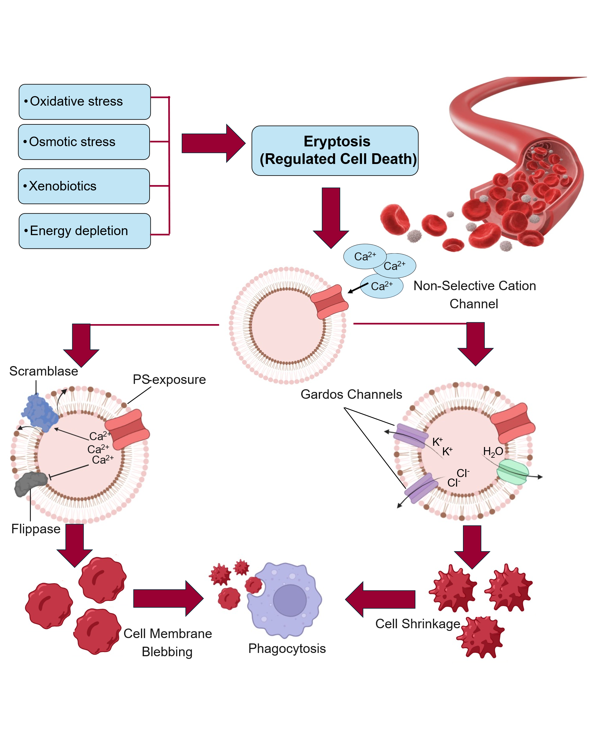
Eryptosis (Fig. 1), in erythrocytes, analogous to apoptosis is characterized through shrinkage of erythrocytes , blebbing of cell membrane and mainly by externalization of (PS) phosphatidylserine on membrane. This process plays critical role in removing damaged, senescent or dysfunctional erythrocytes from blood. By doing so it minimizes the inflammatory responses and prevents hemolysis. Eryptosis is triggered by a various factors like energy depletion, oxidative stress, hyperosmolarity, exposure to heavy metals and xenobiotics. Eryptosis is a tightly regulated process by a complex network of signaling pathways comprising calcium influx, formation of ceramide and caspase-independent mechanisms. Under normal physiological conditions this process is essential for erythrocytes turnover, dysregulated eryptosis may lead to a number of pathological conditions like anemia, cardiovascular diseases or chronic kidney diseases. Hence, maintenance of eryptotic process is crucial for the maintenance of integration of erythrocytes and overall hematological health (Tkachenko et al., 2025).
MECHANISM OF ERYPTOSIS
Eryptosis is a regulated form of erythrocyte death that occurs prior to their natural senescence under both physiological and pathological conditions. This process is clinically significant because it can be induced or accelerated by various factors such as osmotic shock, energy depletion, oxidative stress, exposure to heavy metals, xenobiotic, and antibiotics. Oxidative stress and osmotic shock damage erythrocytes and subsequently harm the lungs and kidneys and various changes occur in erythrocyte membranes such as shrinkage of cell, blebbing of cell membrane and phosphatidylserine exposure on the surface of the cell, which is characteristic of eryptosis and apoptosis (Alghareeb et al., 2023).
Ca+2
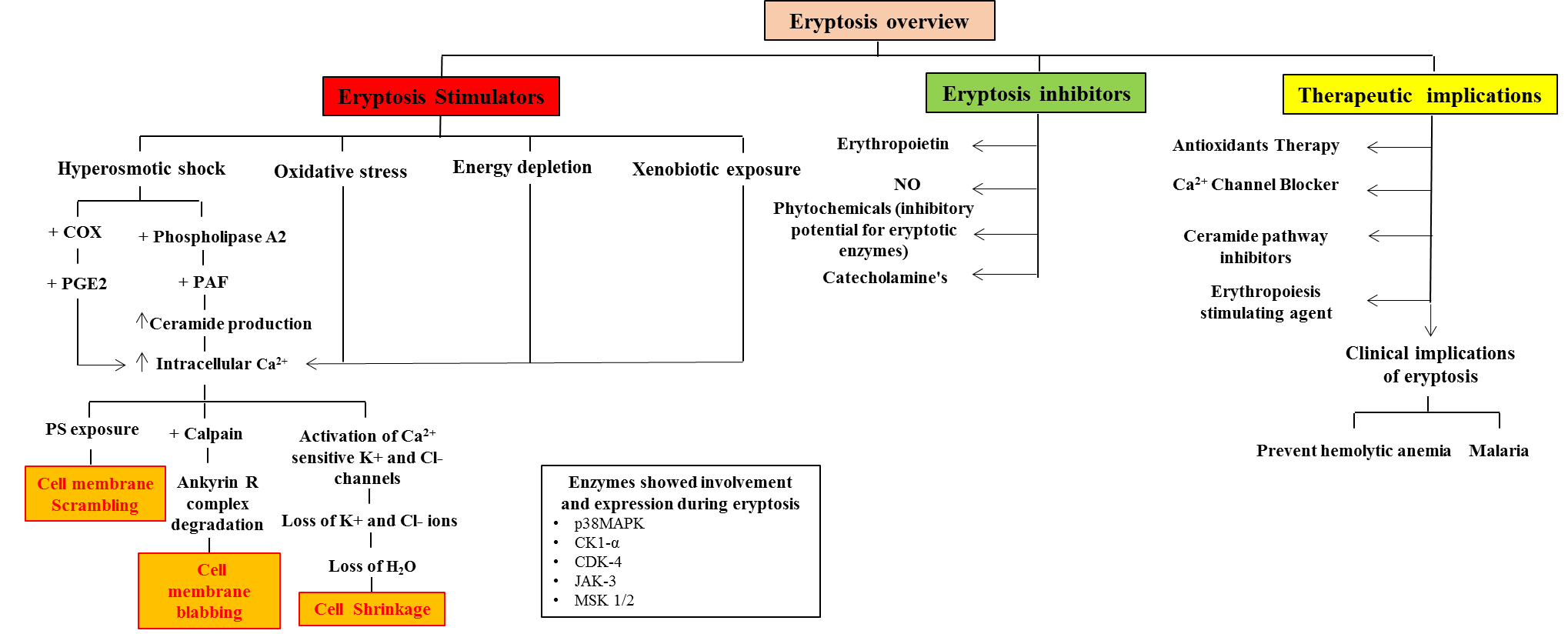
Eryptosis is triggered in various conditions but the major trigger is the increase of intracellular calcium levels due to oxidative stress and hyperosmotic shock which is affected by stimulation of cyclooxygenase with prostaglandins (PGE2) generation which activate cation channel (Pretorius et al., 2016) . Increase in cytosolic Ca+2 activate a non-selective Ca2+ -sensitive K+ channel also called gardos channel which allow influx of Ca+2 and Na+ with subsequent efflux of K+ into erythrocytes and cause hyperpolarization of cell membrane (Lang & Lang, 2015). Activation of calcium permeable channel also exit Cl- due to enhanced electrical gradient, then ultimately cellular loss of KCl and water from erythrocytes. This eventually causes cell shrinkage which is characteristic of eryptosis. leading to shrinkage of cell (Tkachenko et al., 2025). The increase of intracellular Ca2+ also activate Cysteine endopeptidase calcium activated protease which involved in degradation of ankyrin R complex, cytoskeleton of protein consequential increased erythrocytes adhesiveness and cause membrane blebbing.
Due to the loss of Cl- ions there is a release of prostaglandin E2 (PGE2) which also elevates the cytosolic Ca2+ levels and phospholipid scrambling of membrane. This scrambling of phospholipids on membrane involve the exposure of phosphatidylserine from the inner to outer leaflet of the erythrocytes membrane and disrupts the membrane symmetry. The asymmetric organization of the cell membrane plays fundamental role in the functionality of membrane-associated proteins and their interactions with the underlying cytoskeletal network, thereby influencing cellular mechanical stability and deformability of the membrane, also phospholipids disruption on plasma membrane particularly the localization of phosphoinositides and phosphatidylserine (PS) to the cytoplasmic leaflet, which is fundamental in maintaining erythrocytes membrane integrity through regulation of cytoskeletal anchoring, signal transduction pathways, and membrane curvature (Brun et al., 2021). By facilitating enzymatic reaction phospholipid bilayer play a key role in regulating membrane transport mechanisms and initiating signaling cascade pathways essential for cell hemostasis. The complete cascade of intracellular signaling alerts due to phospholipids like PS, which also affect the cytoskeleton and plays chief role in the destruction of premature RBCs. The exposure of phosphatidylserine PS on the erythrocyte membrane is identified by circulating macrophages with specific phosphatidylserine (PS) receptors, which engulf and remove the erythrocyte from the circulation.
Ceramide
Cell death is also associated with the release of (PAF) platelet activating factor which is a strong phospholipid initiator that causes platelet aggregation and dilation of blood vessels and contributes to inflammatory response. PAF also stimulates ceramide release by activating sphingomyelinase, which hydrolyzes sphingomyelin (a plasma membrane lipoprotein) originating from endogenous erythrocytes or exogenous sources. Ceramide is a bioactive lipid involved in inflammatory signaling molecules which is release into the plasma and enhances the activity of calcium-sensitive potassium (K⁺) channels. Osmotic stress can further stimulate PAF release through the activation of phospholipase enzymes. The accumulation of ceramide in the erythrocyte membrane promotes sarcolemmal lipid scrambling, leading to the externalization of phosphatidylserine, a hallmark of eryptosis. This ceramide-mediated transbilayer lipid redistribution may underlie the observed membrane asymmetry loss during erythrocyte stress responses (Alghareeb et al., 2023).
Like apoptosis, eryptosis is also triggered by to various kinase dependent signaling pathways. Exposure to various cell stressors induced eryptosis by regulating signaling cascades involving activation of kinases like p38 MAPK, CK1α, JAK3, PKCα, MSK1/2, and CDK4, which promote Ca²⁺ influx and PS externalization. AMPKα1 and cGK1 serve as inhibitory checkpoints, counteracting stress signals and maintaining erythrocyte stability. Nitric oxide (NO) further suppresses eryptosis by inhibiting Ca²⁺-mediated membrane scrambling and promoting redox balance (Qadri et al., 2017).
PHYSIOLOGICAL FUNCTION OF ERYPTOSIS
Eryptosis show distinct physiological function mainly act as defensive and shorten the lifecycle of erythrocytes that are damaged or impaired through any clinically specific disease or injury to eliminate the erythrocytes from flow of circulation. The mechanism of eryptosis ensure that removal of RBCs from circulation maintaining the hemostasis in blood. In many normal physiological states, spleen maintain the blood permanency through evading premature hemolysis, clearance of senescence and structurally damaged erythrocytes and also eliminating infected cell with pathogens such as harboring Plasmodium falciparum. Along with this it helped in iron recycling, preservation of microcirculation, and regulation of the erythrocyte lifespan, which collectively guarantee efficient oxygen transport. Many genetic disorders such thalassemia sickle cell anemia, and glucose-6-phosphate dehydrogenase deficiency. RBCs particularly more susceptible to energy depletion, osmotic shock, and oxidative stress, their lifespan is shortened to remove defective erythrocytes (Alghareeb et al., 2023).
Eryptosis is exhibited during the erythrocytes natural ageing, which is also characterized by the distinctive increase of Ca2+ level. Aged erythrocytes are more prone to oxidative stress, osmotic shock, and depletion of energy as they may have lost its capability to protect itself from these harmful disorders. Enhanced eryptosis may lead to various clinical complications such as anemia, vascular blockages, oxidative and metabolic imbalances. Overactive or insufficient eryptosis can also enhance blood clotting complications, hemolytic syndromes, and impaired microcirculation, resembling the role of apoptosis. Like apoptosis in nucleated cells, eryptosis happens physiologically to remove damaged and dysfunctional mature erythrocytes from the bloodstream.
The prevention of hemolysis and its associated consequences is a crucial physiological role of eryptosis. When energy depletion occurs, dysregulation of Na+/K+- ATPase and subsequent ion leakage cause hemolysis. This allows water and Na+ and Cl− ions to enter the erythrocytes causing erythrocyte enlargement moreover loss of cellular K+ ions can counterbalance initially influx of Na+ ions. However excessive loss of K+ ions causes membrane depolarization and facilitate entry of Cl− ions. Rupturing of erythrocytes membrane leads to the release of undesirable erythrocyte components including hemoglobin into the circulatory system are caused by the rise in cell volume also manifested as cell swelling. Early removal of stressed erythrocytes prevents the undesired release of hemoglobin (Hb) and damage-associated molecular patterns (DAMPs), both of which can further amplify oxidative damage and inflammation (Tkachenko & Havranek, 2025).
In malaria, elevated rate of incidence of eryptosis is beneficial. Malaria in humans is caused by infection with the Plasmodium protozoan. The mosquito of the genus Anopheles is the arthropod vector that spreads it. Because in erythrocytes Plasmodium falciparum parasite inhibit its growth, the species that parasitize humans are Plasmodium malaria, Plasmodium falciparum, Plasmodium knowlesi, Plasmodium vivax, Plasmodium knowlesi and Plasmodium ovale (Brun et al., 2021). Once the erythrocyte is infected oxidative stress increases and Ca2+ ions enter through the cation channels, causing eryptosis. Ultimately, elevated Ca2+ levels in the erythrocyte triggered mechanism of eryptosis (Fig. 2) leading to removal of infected cell from circulation, espite the parasite reliance on the erythrocyte channel from nutrient uptake (Scovino et al., 2022).
Physiological Adaptation in Newborns
Newborns and Fetus both are distinct type of hemoglobin than grownups since they attain their level of oxygen from the placenta rather than lungs during the process of respiration. Compared to normal hemoglobin, fetal hemoglobin (HbF) has a greater attraction for oxygen. This enables the placental circulation to supply the embryo with adequate oxygen. Because fetal hemoglobin has a greater affinity for oxygen, the fetal erythrocyte's hemoglobin can be partially oxygenated, and less oxygen can be released to the periphery (Nader et al., 2021).
Fetal hemoglobin is not appropriate for efficient exchange of gases after birth; conversely, fetal erythrocytes are more are more resilient to oxidative stress and show high resistance to the elimination of Cl ions level, osmotic shock, platelets activating factor, and prostaglandin E2. Following their exposure to inspired oxygen after birth, these erythrocytes are detached from the neonate's circulation due to the sensitivity of fetal hemoglobin to oxygen. This causes eryptosis, which removes HbF-containing cells from circulation and replaces them with adult-type erythrocytes that are more suited for postnatal gas exchange (Tkachenko et al., 2025).
ERYPTOSIS IN DISEASE
Eryptosis becomes pathogenic when excessively activated or inadequately regulated.
Pathophysiology of Anemia
Anemia is a condition of premature destruction of red blood cells resulting in reduced oxygen supply and compensatory erythropoiesis which results in eryptosis (Usuki, 2015). It is divided into two categories: intrinsic, e.g., membrane defects, enzymopathies, hemoglobinopathies and extrinsic, e.g., autoimmune, microangiopathic, infectious or drug-induced. Common intrinsic causes include G6PD deficiency, hereditary spherocytosis, thalassemia and sickle cell disease. Complement activation, mechanical fragmentation and antibody-mediated lysis are examples of extrinsic causes. Indirect bilirubin, increased lactate dehydrogenase (LDH), reticulocyte count, and decreased haptoglobin which commonly observed in laboratories (Bartolmäs et al., 2018). Chronic disease anemia (ACD) is the reduction of iron utilization and erythropoiesis as a result of chronic inflammation. Inflammatory cytokines cause elevated hepcidin levels, which block iron uptake and sequestration reducing the production of red blood cell (Cheng et al., 2011).
Diabetes Mellitus
Diabetes mellitus (DM) comprises wide variety of metabolic disorders characterized by chronic hyperglycemia and oxidative stress due to reduced insulin secretion. This metabolic disorder contribute to progressive vascular complications such as nephropathy, retinopathy, neuropathy and atherosclerosis (Ogunsanmi et al., 2022). Diabetes associated with decrease red blood cell (RBC) lifespan not because of decreased erythropoiesis but rather to increased eryptosis (Giacco & Brownlee, 2010). Eryptosis in Diabetes is mechanistically associated to glycoxidation whereby oxidative stress chronic hyperglycemia and promote non-enzymatic glycation of membrane proteins leading to the accumulation of (AGEs) advanced glycation end-products such as (CML) carboxymethyl-lysine, (CEL) carboxyethyl-lysine and methylglyoxal (MG) which impairs the shape and functions of red blood cells (Awasthi et al., 2015).These advanced glycation end-products disrupts membrane integrity and impair erythrocytes deformability and enhance phosphatidylserine exposure which is a hallmark of eryptosis. Among AGEs Methylglyoxal play central role inducing oxidative stress and ATP depletion through inhibition of glycolytic enzymes. This impair glutathione synthesis and glutathione peroxidase activity sensitizing erythrocytes to eryptosis. Eryptotic erythrocytes have increased endothelial adhesion and decreased deformability which promotes endothelial dysfunction (ED) and impaired the microvascular. Apart from oxidative mechanism Ca2+independent pathways such as caspase 3 activation also trigger eryptosis in diabetes (Mohammedi et al., 2015).
Chronic Kidney Disease (CKD)
CKD is ongoing and unrecoverable damage in kidney function characterized by a glomerular filtration rate <60 mL/min/1.73 m² for higher than 3 months or after kidney damage (Kimura et al., 2018). A key complication of CKD is renal anemia, primarily due to decreased erythropoietin production and iron deficiency. However, eryptosis play significant role in anemia associated with CKD. Eryptosis exacerbated by factors including oxidative stress, inflammation, loss of energy and uremic toxin accumulation which disrupt the kidney function (Li et al., 2022). This process sets up a vicious cycle of accelerated erythrocytes destruction. Uremic toxins for example urea, indoxyl sulphate, acrolein, indole-3-acetic acid, and p-cresol have been demonstrated to directly trigger eryptosis (Bissinger et al., 2024). Indoxyl sulphate increases cytosolic Ca2+ levels and scrambles erythrocyte membranes leads to increased phosphatidylserine (PS) exposure. It also raises ceramide levels a critical mediator of eryptosis. Furthermore, indoxyl sulphate causes oxidative stress through pathways involving Organic Anion Transporter 2 (OAT2) and NADPH oxidase which are independent of glutathione (GSH). These mechanisms may contribute to the pathophysiology of renal anemia. The combined effect of these molecular abnormalities increases eryptosis and exacerbates anemia in CKD (Butt et al., 2025)
Malaria
Malaria is a vector born disease that occurs due to parasites of zoonotic origin produced by protozoan parasites of the genus Plasmodium. Plasmodium falciparum is the most pathogenic and virulent species (Boulet et al., 2018). It is globally responsible for a majority of malaria-related mortality and morbidity. P. falciparum infection can cause asymptomatic or dangerous clinical conditions such as cerebral malaria and malaria-induced anemia (MIA) which is characterized by a significant decrease in hemoglobin levels (~5 g/dL). Anemia in malaria arises from the combination of impaired erythropoiesis, hemolysis and eryptosis. The destruction of infected and uninfected erythrocytes through eryptosis contributes significantly to MIA and also serve as host defense mechanism by limiting parasites proliferation. High quantities of reactive oxygen species are produced by infected erythrocytes, which increases oxidative stress and activates non-selective cation channels. This facilitates the intracellular influx of Na⁺ and Ca2+. A major cause of eryptosis is elevated cytosolic Ca2+ which is indicated by the externalization of phosphatidylserine a signal for macrophage-mediated phagocytosis. Activated immune cells produce more reactive oxygen species and reactive nitrogen species which promotes erythrocytes destruction. Genetic traits that predispose erythrocytes to eryptosis such as sickle cell trait, β-thalassemia trait, iron deficit, G6PD deficiency and impaired transporters including GLUT1 and AE1transpoter give partial protection against severe P. falciparum infection by promoting faster clearance of infected erythrocytes (Scovino et al., 2022).
Sepsis and Inflammation
Sepsis is a complicated clinical disease characterized by systemic inflammation, immunological dysregulation and multiorgan failure in response to infection. It is related with a significant morbidity, death, and healthcare burden (Singer et al., 2016). The pathogenesis includes oxidative stress, immunological imbalance, endothelial dysfunction, coagulation problems and significant cellular damage. Sepsis causes a systemic inflammatory response with high production of cytokines which is pro-inflammatory cytokines e.g., IL-6, TNF-α, IL-1β. These cytokines coupled with (PAMPs) pathogen-associated molecular patterns and (DAMPs) damage-associated molecular patterns (DAMPs) stimulate immune cells resulting in increased production of reactive oxygen species and reactive nitrogen species. The outcome of oxidative stress damages erythrocyte membranes and disrupts ion homeostasis (Yu et al., 2020). The oxidative injury and cytokine signaling induce the opening of non-selective cation channels which allowing an influx of Ca²⁺ into erythrocytes. Increased intracellular calcium stimulates scramblase while inhibiting flippase resulting in phosphatidylserine (PS) externalisation on the erythrocyte surface a characteristic of eryptosis. PS exposure marks erythrocytes for identification and removal by macrophages thereby leading to anemia in sepsis (Franco et al., 2019).
DETECTION AND MEASUREMENT OF ERYPTOSIS
Laboratory assessment of eryptosis includes;
Flow cytometry with Annexin V-FITC for PS exposure
Annexin V-FITC is a widely used method for detection of phosphatidylserine (PS) exposure on the cell surface, an indication of apoptosis (programmed cell death). Annexin V, is a binding protein that bind phospholipid, and also has a strong calcium-dependent attraction of annexin V for phosphatidylserine (PS) (Demchenko, 2013).
Detection methodology: Annexin V-FITC conjugates used in flow cytometry to detect PS exposure on outer leaf let of cell membrane. It can detect PS on outer membrane of early apoptotic cells (Alshehade et al., 2024). It can distinguished between cells showing V positive and propidium iodide negative (Annexin V+/PI−) for early apoptosis and late apoptosis (Annexin V+/PI+).
Advantages: One of the major advantage of this technique is its specificity and high sensitivity. It is used for quantitatively investigation of apoptotic cell population. It is also Applicable for different cell types and species (Saxena et al., 2024).
Limitations: However this method also has some limitations. It requires calcium ions for binding of Annexin-V. In addition, possible false positives in cells that are being activated or differentiated. Furthermore, not every apoptotic pathway involve PS exposure, so that’s why it may limit the assay’s ability for detection (Kato, 2025).
Ca2+ imaging using calcium-sensitive fluorescent dyes
A variety of cellular functions, such as muscular contraction, neurotransmitter release, gene expression, and cell death, are mediated by calcium ions (Ca2+). Physiological and pathological cellular processes can be better understood by tracking intracellular calcium dynamics (Pikor et al., 2024).
Detection Methodology: A common method to visualize and quantify changes in intracellular calcium concentrations (Ca²), is calcium imaging with calcium-sensitive fluorescent dyes (Bootman et al., 2013). Fluorescence intensity of these dyes increases as they bind to Ca2+ , and excitation ratio or fluorescence emission varies as a result of Ca2+ binding (Sheng et al., 2024).Use of calcium-sensitive fluorescent dyes to load cells (such as Fura-2 AM, Calcium Green, and Indo-1 for ratiometric imaging or Fluo-4 AM for single-wavelength intensity imaging) (Bootman & Bultynck, 2020). It can detect variations in intracellular Ca2+ concentrations (such as calcium transients, sparks, or oscillations) that indicate signaling or cell activation. Numerous cellular functions, including muscular contraction, cell death (apoptosis), and neuronal activity (Pikor et al., 2024).
Advantages: There are several advantages of these dyes used in this method. This method provide a high temporal resolution for dynamic processes. These dyes are compatible with many cell types. Furthermore, these fluorescent markers are useful for the quantitative evaluation of acute intracellular Ca2+ (Sheng et al., 2024)
Limitations: This technique has many advantages but also several limitations are associated with this method, as some dyes are saturate or unable to resolve very quick or tiny amplitude Ca2+ signals. Dye buffering may change calcium dynamics. Non-ratiometric dyes are susceptible to loading fluctuations. There is a chance of photobleaching and phototoxicity during imaging (Zhou et al., 2021).
ROS Detection via DCFDA Staining
Reactive oxygen species (ROS) are oxygen-containing molecules that are chemically reactive, including hydroxyl radicals, hydrogen peroxide, and superoxide anion. ROS are vital for cell signaling and homeostasis, but at high concentration, can lead to oxidative stress, which causes damage to cells and involved in a number of clinical disorders, such as inflammation, cancer, and neurodegeneration (Ghosh et al., 2018). The most popular method for directly determining a cell's redox state is (DCFH-DA) 2′-7′-Dichlorodihydrofluorescein diacetate (Figueroa et al., 2018). Various diseases in human, like diabetes, atherosclerosis, cancer, and other neurological conditions, are significantly influenced by ROS and oxidative damage. Therefore, it is necessary to measure the capacity of ROS and the oxidative damage they produce.
Detection Methodology: After exposing live cells to H₂DCFDA (~1–10 µM) for around 30 minutes in the dark, wash the cells and promptly examine their fluorescence (such as the FL-1 channel) using flow cytometry, microscopy, or a plate reader. To quantify intracellular ROS generation, DCFDA (H₂DCFDA) staining require loading live cells with the non-fluorescent dye (Yoon et al., 2018).
Advantages: This is very simple and inexpensive method with high sensitivity to redox state changes. Compatible with flow cytometry, plate readers, and microscopy. It is an easy way to detect ROS because direct imaging of ROS in biological sample is difficult (Zhang et al., 2025).
Limitations: This technique also present some limitations. It lack of specificity and it is unable to differentiate between different forms of ROS. In addition, Dye is susceptible to auto-oxidation, photobleaching, and background Fluorescence in phenol-red or conditions. Signals are dependent on cell type, dye uptake, and esterase activity (Kato, 2025)
Microscopic Analysis for Morphological Changes
Cell microscopy is a common method that is probably used in laboratories that works with cells by using fluorescent markers and various visual assay. Examining tissues and cells at the microscopic level in order to detect and describe changes in their size, shape or structure is known as microscopic analysis for morphological changes. This method is frequently used to diagnose illnesses or investigate cellular functions (Quadri et al., 2024).
Detection Methodology: Electron microscopy (TEM/SEM) or light microscopy (phase-contrast or DIC), optionally with H&E or fluorescent nuclear stains (e.g., DAPI, AO) (Czerwińska-Główka & Krukiewicz, 2021). It can detect cell shrinkage, cytoplasm densification, chromatin condensation/pyknosis, membrane blebbing, and the production of apoptotic bodies or nuclear fragmentation (Mandelkow et al., 2017).
Advantages: Light microscopy is affordable technique, and capable of real-time imaging. Electron microscopy provides excellent specificity for apoptotic morphology and ultrastructure detail at high resolution. It is very cost-effective and also it offer direct visual confirmation of cell death stages (Cibreiros et al., 2025)
Limitations: Despite its several advantages, it also exhibit a lot of limitations like, poor quantification, subjective and operator-dependent; lengthy sample preparation; limited throughput, destructive, and mostly detecting late apoptotic stages. Electron microscopy may miss early alterations and varies by cell type (McCafferty et al., 2024).
Ceramide Quantification Using Immunostaining or Mass Spectrometry
Mass spectrometry (MS) is an analytical method to measure mass-to-charge ratio and immunostaining is a laboratory technique uses antibodies to detect molecules like lipids. These both can be used to quantify ceramide; each has unique benefits and drawbacks based on the sample type, sensitivity needs, and experimental objectives. These both involve either in extracting lipids for LC-ESI-MS/MS with isotopically labeled ceramide standards. These both techniques has unique benefits as well as drawbacks based on the sample type, sensitivity and experimental objectives.
Detection Methodology: immunostaining is a method to visualize ceramide localization in fixed cells or tissues which employs anti-ceramide antibodies for microscopy (Greene et al., 2023). Cellular and subcellular location of endogenous ceramide can be detected through this procedure. It can find out the absolute levels of ceramide species.
Advantages: Ceramide in cells or tissues can be seen by using this approach, which employ antibodies. Ceramide can co-localize with proteins or organelles and be seen in situ. It is comparatively inexpensive and easily available (Greene et al., 2023).
Limitations: There are also some drawbacks linked to this approach. It is a semi-quantitative method and antibody specificity (cross-reactivity, epitope masking) is still a major concern. Artifacts caused by fixation and uneven staining in different samples (Canals & Hannun, 2024)
LC-ESI-MS/ MS
Very sensitive and precise analytical method for measuring ceramide. It offers precise measurement of ceramide species (Begou et al., 2021). Absolute concentrations of each ceramide molecular species in extracted biosamples (chain length, saturation) (Wang et al., 2021)
Advantages: It is highly sensitive and specific technique. It can measure many ceramide species in a single run. It has robust validation: linear range (pg–ng), accuracy, and repeatability verified in tissues and plasma.
Limitations: This procedure also has several limitations. It does not show spatial or cell-level resolution. Only bulk analysis is possible. Additionally, it requires complex extraction. In this method, ion suppression and matrix effects can occur compromising the accuracy of results. Moreover, costly equipment and technical know-how are needed (Wang et al., 2021).
THERAPEUTIC IMPLICATIONS
Eryptosis shows significant role in maintaining erythrocyte quality and preventing the circulation of infected cells. However, unregulated eryptosis may contribute to certain pathological conditions, including anemia, thrombosis, and infections (Alghareeb et al., 2023). Therefore, by either inhibiting excessive eryptosis or selectively inducing it, targeted therapies can be tailored to treat Certain conditions like chronic kidney disease, cancer, malaria, and sepsis. Consequently, modulation of eryptosis pathways offers a promising approach for improving outcomes in both hematologic and systemic diseases.
Antioxidants Therapy
Oxidative stress is one of the major triggers of eryptosis (Tkachenko & Havránek, 2023) Therefore, antioxidant therapy can be a promising approach for controlling eryptosis and its downstream complications. This therapeutic approach helps restore redox homeostasis by supporting the endogenous defense system, stabilizing the plasma membrane, inhibiting oxidative signaling pathways, and, most importantly, scavenging free radicals. Naturally antioxidants, known as endogenous antioxidants, like (GPx) glutathione peroxidase, (SOD) superoxide dismutase and (CAT) catalase scavenge free radicals within cells. In addition, antioxidants can be administered externally and are referred to as exogenous or nutraceutical antioxidants like N-acetylcysteine (NAC), which donates a thiol group (Ran et al., 2015); polyphenols such as resveratrol and quercetin, which inhibit oxidative signaling pathways including NF-κB and p38 MAPK; and vitamins like vitamin C and vitamin E. Furthermore, synthetic antioxidants have been developed for therapeutic use and are classified as pharmacological antioxidants. One such example is edaravone, a potent free radical scavenger used to counter oxidative stress in clinical settings.
Calcium Channel Blockers
Non-selective cation channels such as voltage-independent stretch-activated channels, mechanosensitive Piezo1 channels, and Transient Receptor Potential Canonical channels (TRPC6), are involved in Ca²⁺ influx (Michelucci & Catacuzzeno, 2024). Elevated cytosolic Ca²⁺ levels activate scramblases and calpains, leading to phosphatidylserine (PS) externalization and cytoskeletal degradation, respectively. Additionally, increased cytosolic Ca²⁺ activates Grados channels leading to K⁺ leakage and ultimately cell shrinkage (Yan et al., 2024). Certain calcium channel blockers are known to inhibit these non-selective cation channels, thereby inhibiting eryptosis, such as verapamil, nifedipine, and amlodipine.
Ceramide Pathway Inhibitors
Ceramide is a sphingolipid metabolite that, when elevated in erythrocyte membranes, promotes phosphatidylserine (PS) externalization. It disrupts membrane symmetry and activates calcium-permeable cation channels, leading to calcium influx and the induce eryptosis (Virzì et al., 2025). Therefore, targeting the ceramide signaling and biosynthesis pathways may represent a novel therapeutic strategy to modulate eryptosis. In erythrocytes, ceramide is produced through the sphingomyelinase pathway, wherein sphingomyelinase hydrolyzes membrane sphingomyelin (Bettioui et al., 2023). Sphingomyelinase inhibitors such as GW4869 and desipramine (Kho et al., 2022) may be employed to inhibit the ceramide pathway.
Erythropoiesis Stimulating Agents
(ESAs) is a Erythropoiesis-stimulating agents (ESAs) belong to a class of drugs that mimic the action of erythropoietin (EPO), an endogenous glycoprotein hormone that plays a vital role in erythropoiesis. EPO favors survival, differentiation, and proliferation of erythroid progenitor cells and enhances erythrocyte production in the bone marrow. Certain ESAs activate the JAK2/STAT5 signaling pathway through PKC, PI3K, and MAPK cascades, which are crucial for erythropoiesis (Tóthová et al., 2021). In addition to their erythropoietic function, ESAs are known for their cytoprotective properties, possessing antioxidant potential and reducing oxidative stress, thereby promoting cell survival. ESAs also directly mediate anti-eryptotic pathways via MAPK signaling (Table 1), as EPO receptors are expressed on mature erythrocytes. Commonly used ESAs include epoetin alfa, a recombinant human erythropoietin (Anand et al., 2021); darbepoetin alfa, a modified form of erythropoietin with a prolonged half-life (Qian, 2025); and methoxy polyethylene glycol-epoetin beta, which activates the erythropoietin receptor. Additionally, next-generation ESAs such as HIF-PH inhibitors have emerged, which stimulate endogenous EPO production by stabilizing hypoxia-inducible factors (Odawara et al., 2024).
| Molecular signaling /pathway | Functional role | Activated by stress condition | Mechanistic contribution to eryptosis | References | ||
|---|---|---|---|---|---|---|
| p38 MAPK (p38 Mitogen-Activated Protein Kinase) | Stress-activated pro-eryptotic kinase | Oxidative stress, osmotic shock, inflammatory cytokines | Activates stress signaling → promotes Ca²⁺ influx and PS externalization | Restivo et al., 2022 | ||
| CK1α (Casein kinase 1 alpha) | stress-responsive pro-eryptotic kinase | Oxidative/osmotic stress, energy depletion | Phosphorylates cytoskeletal/membrane proteins → enhances Ca²⁺ entry, scramblase activation, PS exposure | Alfhili et al., 2024 | ||
| JAK3 (Janus kinase 3) | Cytokine-mediated pro-eryptotic regulator | Glucose/energy depletion, cytokine stimulation | Phosphorylation during energy depletion enhances PS exposure; inhibition blunts eryptosis | Tkachenko & Havranek, 2025 | ||
| PKCα (Protein kinase c alpha) | Membrane scrambling inducer | Oxidative stress, osmotic stress, phorbol esters | Activates Ca²⁺ entry and scramblase → PS exposure | Alfhili et al., 2024 | ||
| CDK4 (Cyclin dependent kinase 4) | Cell cycle–linked pro-eryptotic regulator | Oxidative stress condition | Links cell cycle–related signaling to eryptosis induction | Lang et al., 2015a | ||
| MSK1/2 (Mitogen and stress activated kinase ) | Downstream MAPK pathway mediators |
| Mediate MAPK cascades contributing to eryptosis | Lang et al., 2015b | ||
| AMPKα1 (AMP-activated Protein Kinase alpha 1) | Metabolic checkpoint, anti-eryptotic regulator | Energy depletion (activated as protective response) | Counteracts stress signals, maintains erythrocyte stability | Restivo et al., 2022 | ||
| cGK 1 (Cyclic GMP-dependent Protein Kinase 1) | cGMP-mediated anti-eryptotic regulator | cGMP signaling, nitric oxide pathway | Suppresses stress-induced eryptosis and stabilizes cell membrane | Repsold & Joubert, 2018 | ||
| NO (Nitric oxide) | anti-eryptotic mediator | Oxidative stress, hypoxia, inflammation | Inhibits Ca²⁺-mediated scrambling; maintains redox balance | Repsold & Joubert, 2018 |
Abbreviations: TRPC, transient receptor potential; SERCA, sarco/endoplasmic reticulum Ca²⁺-ATPase; SOCE, store- operated calcium entry; MAPK, mitogen activated protein kinase; MKK3/6 MAPK, Kinase 3 and 6; ATP, adenosine triphosphate; HIF-1 α, hypoxia-Inducible factor 1-alpha; ROS, reactive oxygen species
Targeted Eryptosis Induction
Some pathogens directly exploit erythrocytes such as the malaria parasite Plasmodium resides within these cells and propagates intracellularly. Under such conditions, the clearance of defective erythrocytes from the circulatory system may serve as a potential therapeutic strategy to eliminate infected cells. Ultimately, to remove dysfunctional erythrocytes, eryptosis can be induced through certain pathways such as calcium influx, oxidative stress, ceramide formation, energy depletion, and the activation of different signaling kinases such as p38 MAPK, CK1α, PKC, JNK, and AMPK. TRPC Transient Receptor Potential; SERCA Sarco/Endoplasmic Reticulum Ca²⁺-ATPase; SOCE Store-operated calcium entry; MAPK Mitogen activated protein kinase; MKK3/6 MAPK Kinase 3 and 6; ATP Adenosine triphosphate; HIF-1 α Hypoxia-Inducible Factor 1-alpha; ROS Reactive Oxygen Species.
References
- Alfhili MA, SA Alghareeb, GA Alotaibi et al., 2024. Galangin triggers eryptosis and hemolysis through Ca2+ nucleation and metabolic collapse mediated by PKC/CK1α/COX/p38/Rac1 signaling axis. International Journal of Molecular Sciences 25:12267.
- Alghareeb SA, MA Alfhili & S Fatima, 2023. Molecular mechanisms and pathophysiological significance of eryptosis. International Journal of Molecular Sciences 24:5079.
- Alshehade SA, HA Almoustafa, MA Alshawsh et al., 2024. Flow cytometry-based quantitative analysis of cellular protein expression in apoptosis subpopulations: A protocol. Heliyon 10:33665.
- Anand S, J Al-Mondhiry, K Fischer et al., 2021. Epoetin alfa-epbx: a new entrant into a crowded market: a historical review of the role of erythropoietin stimulating agents and the development of the first epoetin biosimilar in the United States. Expert Review of Clinical Pharmacology 14:1-8.
- Awasthi S, S Gayathiri, R Ramya et al., 2015. Advanced glycation-modified human serum albumin evokes alterations in membrane and eryptosis in erythrocytes. Applied Biochemistry and Biotechnology 177:1013-24.
- Bartolmäs T, B Mayer, AH Balola et al., 2018. Eryptosis in autoimmune haemolytic anaemia. European Journal of Haematology 100:36-44.
- Begou OA, O Deda, E Karagiannidis et al., 2021. Development and validation of a RPLC-MS/MS method for the quantification of ceramides in human serum. Journal of Chromatography B 1175:122734.
- Bettioui T, C Chipeaux, KB Arfa et al., 2023. Development of a new online SPE-HPLC-MS/MS method for the profiling and quantification of sphingolipids and phospholipids in red blood cells-Application to the study of Gaucher's disease. Analytica Chimica Acta 1278:341719.
- Bissinger R, SM Qadri & F Artunc, 2024. Eryptosis: a driver of anemia in chronic kidney disease. Current Opinion in Nephrology and Hypertension 33:220-5.
- Bootman MD & G Bultynck, 2020. Fundamentals of cellular calcium signaling: a primer. Cold Spring Harbor Perspectives in Biology 12:a038802.
- Bootman MD, K Rietdorf, T Collins et al., 2013. Ca2+-sensitive fluorescent dyes and intracellular Ca2+ imaging. Cold Spring Harbor Protocols 2013:83-99.
- Boulet C, CD Doerig & TG Carvalho, 2018. Manipulating eryptosis of human red blood cells: a novel antimalarial strategy? Frontiers in Cellular and Infection Microbiology 8:419.
- Brun J-F, E Varlet-Marie, J Myzia et al., 2021. Metabolic influences modulating erythrocyte deformability and eryptosis. Metabolites 12:4.
- Butt N, S Nasir, U Ahmad et al., 2025. Curcumin attenuates hippuric acid-induced eryptosis by enhancing antioxidant enzyme activity. Indus Journal of Bioscience Research 3:139-147.
- Canals D & YA Hannun, 2024. Biological function, topology, and quantification of plasma membrane ceramide. Advances in Biological Regulation 91:101009.
- Cheng P-P, X-Y Jiao, X-H Wang et al., 2011. Hepcidin expression in anemia of chronic disease and concomitant iron-deficiency anemia. Clinical and Experimental Medicine 11:33-42.
- Cibreiros MP, MHC Leão, C Mermelstein et al., 2025. Show me the image: a systematic analysis on how results are represented in publications from different fields of biomedical and biological research. Anais da Academia Brasileira de Ciências 97:e20241023.
- Czerwińska-Główka D & K Krukiewicz, 2021. Guidelines for a morphometric analysis of prokaryotic and eukaryotic cells by scanning electron microscopy. Cells 10:3304.
- Demchenko AP, 2013. Beyond annexin V: fluorescence response of cellular membranes to apoptosis. Cytotechnology 65:157-172.
- Figueroa D, M Asaduzzaman & F Young, 2018. Real time monitoring and quantification of reactive oxygen species in breast cancer cell line MCF-7 by 2′,7′-dichlorofluorescin diacetate (DCFDA) assay. Journal of Pharmacological and Toxicological Methods 94:26-33.
- Föller M & F Lang, 2020. Ion transport in eryptosis, the suicidal death of erythrocytes. Frontiers in Cell and Developmental Biology 8:597.
- Franco DM, I Arevalo-Rodriguez, IFM Roqué et al., 2019. Plasma interleukin-6 concentration for the diagnosis of sepsis in critically ill adults. Cochrane Database Syst Rev 4:11811.
- Giacco F & M Brownlee, 2010. Oxidative stress and diabetic complications. Circulation Research 107:1058-70.
- Greene M, MJ Hernandez-Corbacho, AG Ostermeyer-Fay et al., 2023. A simple, highly sensitive, and facile method to quantify ceramide at the plasma membrane. Journal of Lipid Research 64:100322.
- Kato TA, 2025. DCFDA assay for oxidative stress measurement in fluorometer. In: DNA Damage Detection: Methods and Protocols (Kato TA, ed.): Springer, USA.
- Kho AR, BY Choi, SH Lee et al., 2022. Administration of an acidic sphingomyelinase (ASMase) inhibitor, imipramine, reduces hypoglycemia-induced hippocampal neuronal death. Cells 11:667.
- Kimura K, T Hosoya, S Uchida et al., 2018. Febuxostat therapy for patients with stage 3 CKD and asymptomatic hyperuricemia: a randomized trial. American Journal of Kidney Diseases 72:798-810.
- Lang E & F Lang, 2015. Triggers, inhibitors, mechanisms, and significance of eryptosis: the suicidal erythrocyte death. BioMed Research International 2015:513518.
- Lang E, C Zelenak, M Eberhard et al., 2015b. Impact of cyclin-dependent kinase CDK4 inhibition on eryptosis. Cellular Physiology and Biochemistry 37:1178-86.
- Lang E, R Bissinger, A Fajol et al., 2015a. Accelerated apoptotic death and in vivo turnover of erythrocytes in mice lacking functional mitogen-and stress-activated kinase MSK1/2. Scientific Reports 5:17316.
- Li D, X Zheng, Y Zhang, X Li et al., 2022. What should be responsible for eryptosis in chronic kidney disease? Kidney and Blood Pressure Research 47:375-90.
- Liu H, Y Li, Y Wang et al., 2025. Red blood cells-derived components as biomimetic functional materials: matching versatile delivery strategies based on structure and function. Bioactive Materials 47:481-501.
- Mandelkow R, D Guembel, H Ahrend et al., 2017. Detection and quantification of nuclear morphology changes in apoptotic cells by fluorescence microscopy and subsequent analysis of visualized fluorescent signals. Anticancer Research 37:2239-44.
- McCafferty CL, S Klumpe, RE Amaro et al., 2024. Integrating cellular electron microscopy with multimodal data to explore biology across space and time. Cell 187:563-84.
- Michelucci A & L Catacuzzeno, 2024. Piezo1, the new actor in cell volume regulation. Pflügers Archiv-European Journal of Physiology 476:1023-39.
- Mohammedi K, N Bellili-Muñoz, SL Marklund et al., 2015. Plasma extracellular superoxide dismutase concentration, allelic variations in the SOD3 gene and risk of myocardial infarction and all-cause mortality in people with type 1 and type 2 diabetes. Cardiovascular Diabetology 14:845.
- Nader E, N Conran, M Romana et al., 2021. Vasculopathy in sickle cell disease: from red blood cell sickling to vascular dysfunction. Comprehensive Physiology 11:1785-803.
- Odawara M, H Nishi & M Nangaku, 2024. A spotlight on using HIF-PH inhibitors in renal anemia. Expert Opinion on Pharmacotherapy 25:1291-9.
- Ogunsanmi AO, T Raheem & WS Adio, 2022. Molecular aspect of diabetes melitus. World Scientific News 170:32-45.
- Pikor D, M Hurła, B Słowikowski et al., 2024. Calcium ions in the physiology and pathology of the central nervous system. International Journal of Molecular Sciences 25:13133.
- Pretini V, MH Koenen, L Kaestner et al., 2019. Red blood cells: chasing interactions. Frontiers in Physiology 10:945.
- Pretorius E, JN Du Plooy & J Bester, 2016. A comprehensive review on eryptosis. Cellular Physiology and Biochemistry 39:1977-2000.
- Qadri SM, R Bissinger, Z Solh et al., 2017. Eryptosis in health and disease: a paradigm shift towards understanding the (patho) physiological implications of programmed cell death of erythrocytes. Blood Reviews 31:349-61.
- Qian H, 2025. Retrospective analysis comparing the effectiveness of epoetin alfa and darbepoetin alfa in cancer related and chronic kidney disease related anemia patients. MedScien 1:1.
- Quadri F, M Govindaraj, S Soman et al., 2024. Uncovering hidden treasures: mapping morphological changes in the differentiation of human mesenchymal stem cells to osteoblasts using deep learning. Micron 178:103581.
- Ran Q, Y Xiang, Y Liu et al., 2015. Eryptosis indices as a novel predictive parameter for biocompatibility of Fe3O4 magnetic nanoparticles on erythrocytes. Scientific Reports 5:16209.
- Repsold L & AM Joubert, 2018. Eryptosis: an erythrocyte's suicidal type of cell death. BioMed Research International 2018:9405617.
- Restivo I, A Attanzio, IC Giardina et al., 2022. Cigarette smoke extract induces p38 MAPK-initiated, fas-mediated eryptosis. International Journal of Molecular Sciences 23:14730.
- Saxena C, V Wadhwan, V Kumar et al., 2024. A quantitative assessment of apoptotic index with varying histological degree of dysplasia and squamous cell carcinoma of oral cavity-A light microscopic study. Indian Journal of Dental Research 35:201-5.
- Scovino AM, PRR Totino & A Morrot, 2022. Eryptosis as a new insight in malaria pathogenesis. Frontiers in Immunology 13:855795.
- Sheng C-Q, S-S Wu, Y-K Cheng et al., 2024. Comprehensive review of indicators and techniques for optical mapping of intracellular calcium ions. Cerebral Cortex 34:bhae346.
- Singer M, CS Deutschman, CW Seymour et al., 2016. The third international consensus definitions for sepsis and septic shock (Sepsis-3). Jama 315:801-10.
- Tkachenko A & O Havránek, 2023. Redox status of erythrocytes as an important factor in eryptosis and erythronecroptosis. Folia Biologica 69:116-26.
- Tkachenko A & O Havranek, 2025. Cell death signaling in human erythron: erythrocytes lose the complexity of cell death machinery upon maturation. Apoptosis 30:652-73.
- Tkachenko A, MA Alfhili, J Alsughayyir et al., 2025. Current understanding of eryptosis: mechanisms, physiological functions, role in disease, pharmacological applications, and nomenclature recommendations. Cell Death and Disease 16:467.
- Tkachenko AJA, 2024. Apoptosis and eryptosis: similarities and differences. Apoptosis 29:482-502.
- Tóthová Z, J Tomc, N Debeljak et al., 2021. STAT5 as a key protein of erythropoietin signalization. International Journal of Molecular Sciences 22:7109.
- Usuki K, 2015. Anemia: from basic knowledge to up-to-date treatment. Topic: IV. Hemolytic anemia: diagnosis and treatment. The Journal of the Japanese Society of Internal Medicine 104:1389-96.
- Virzì GM, A Clementi, C Ronco et al., 2025. Red cell death in renal disease: the role of eryptosis in CKD and dialysis patients. Cells 14:967.
- Wang D, P Xu & C Mesaros, 2021. Analytical considerations for reducing the matrix effect for the sphingolipidome quantification in whole blood. Bioanalysis 13:1037-49.
- Yan W, R Wu, Y Lee et al., 2024. Perturbation of calcium homeostasis invokes eryptosis-like cell death in enucleated bone marrow stem cells. Biochemistry and Cell Biology 103:1-11.
- Yoon DS, M-H Lee & DS Cha, 2018. Measurement of intracellular ROS in Caenorhabditis elegans using 2′,7′-dichlorodihydrofluorescein diacetate. Bio-protocol 8:e2774.
- Yu H, Y Liu, M Wang et al., 2020. Myeloperoxidase instigates proinflammatory responses in a cecal ligation and puncture rat model of sepsis. American Journal of Physiology-Heart and Circulatory Physiology 319:H705-21.
- Zhang Y, Y Yi, Y Shu et al., 2025. TRP channels and breast cancer: the role of TRPs in the pathophysiological development. Frontiers in Molecular Biosciences 12:1528663.
- Zhou X, KJ Belavek & EW Miller, 2021. Origins of Ca2+ imaging with fluorescent indicators. Biochemistry 60:3547-54.