

INTRODUCTION
Histopathology is the foundation of disease diagnosis, research, and also involves in the study of tissue sections by using a microscope to determine the presence of pathological changes (Gupta, E et al., 2009). Although it is very important, traditional histopathological examination is so dependent on manual interpretation that may prove to be subjective and time-consuming (Dika et al., 2022). The increasing amount of biopsy samples and the overall lack of trained pathologists worldwide have resulted in an acute necessity for computational tools, which would be useful in the correct analysis, reproducibility, and high-throughput of tissues (Fig. 1).
Machine learning (ML) and deep learning (DL) are known as Artificial intelligence (AI), which has been quickly developed as a solution to these challenges. With the ability to address digital histopathological data with impressive accuracy, AI systems can imitate human mechanisms of thinking and learn complicated patterns using tremendous amounts of data (Bahadir et al., 2024).
Already, AI has proven to be better or equal to human specialists in the detection of breast and lung, prostate and colorectal cancers, and in identifying finer morphological details that might be missed during human examination. Implementation of AI in histopathology is a paradigm shift from qualitative to quantitative assessment of tissues. It makes possible the extraction of high-dimensional features, sometimes called pathomics, that intermediate the microscopic morphology to molecular and clinical outcome (Balestrieri et al., 2025). This chapter will discuss the most important concepts, uses, issues, and future of AI in histopathological image analysis, emphasizing its presence in the future of diagnostic-level pathology.
BACKGROUND OF HISTOPATHOLOGICAL IMAGE ANALYSIS
Overview of Histopathology
Histopathology is the study of thin tissue, which has been stained by using dyes like hematoxylin and eosin (H&E), which help in visualizing cellular and extracellular structures. Pathologists determine features of architecture, cell morphology, and staining patterns to detect disease processes (Lowe et al., 2023). Conventionally, this has been done through manual microscopy that, though proven to be effective, has limitations of variability in its observers and poor reproducibility.
Importance of Digital Pathology
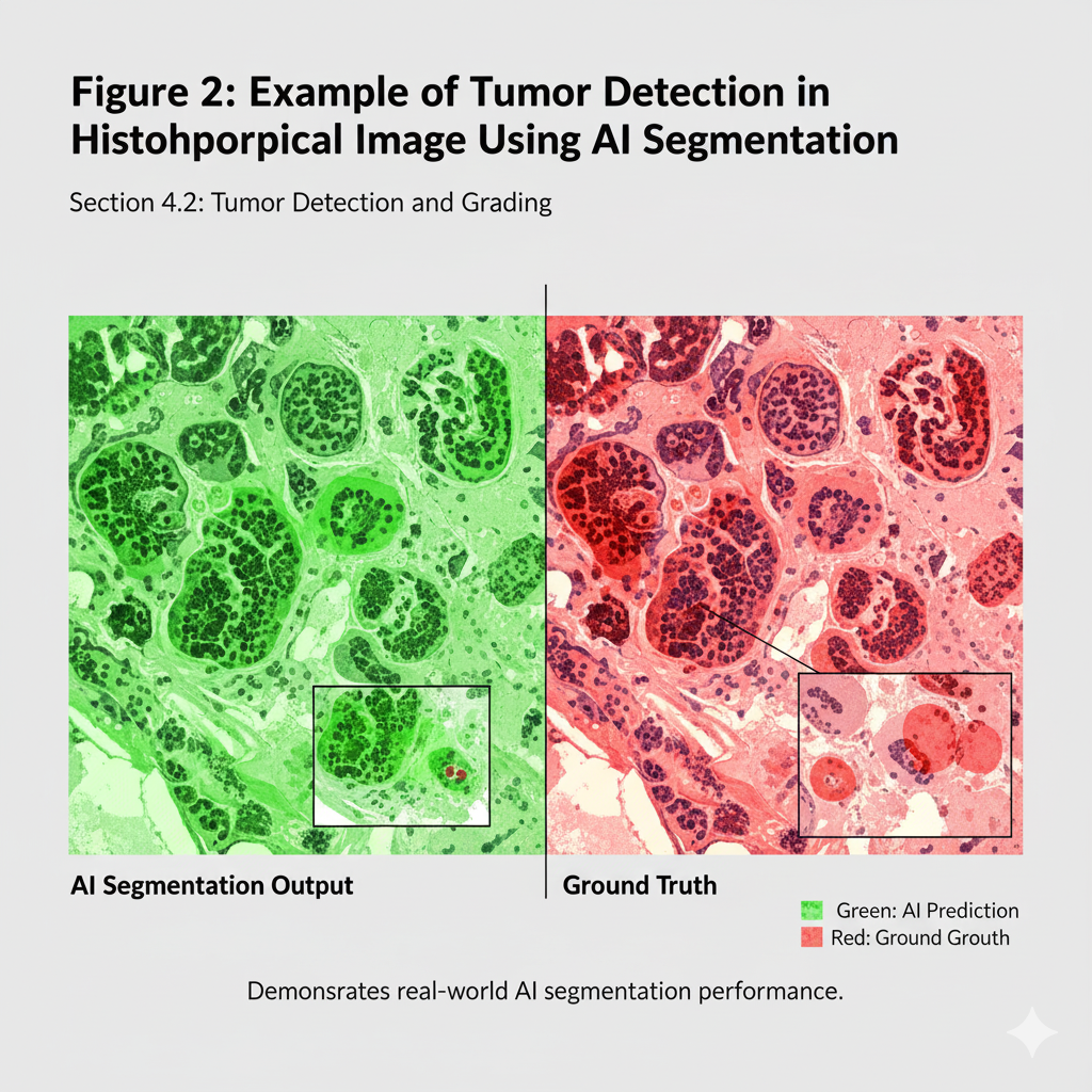
Digital pathology can be defined as the obtaining, storage, distribution and interpretation of pathology data within a digital framework. Whole-slide imaging (WSI) enables the use of glass slides at high resolution, and the resultant digital images can be viewed, annotated, and analyzed by computer (Jain et al., 2024). The digitalization of slides has led to AI usage, which makes it possible to use large-scale data to learn deep learning algorithms (Fig. 2).
Limitations of Conventional Histopathology
Conventional histopathology can be characterized by inter-observer variability, errors caused by fatigue and the inability to quantify the morphological patterns. Besides, complex molecular or prognostic data cannot be readily incorporated into manual examination (Biswas et al., 2025). AI fails to overcome these limitations as it standardizes image perception, quantifies tissue responses, and uncovers possibly hidden patterns that are unseen by humans.
FUNDAMENTALS OF AI AND DEEP LEARNING
Machine Learning in Image Analysis
Machine learning is a method of learning from data in order to make predictions or classifications. Histopathology Traditional ML methods, including support vector machines (SVM), random forests, and k-nearest neighbors, have been used to extract handcrafted features, including cell size, cell shape, cell texture and cell color intensity. Nevertheless, these approaches require feature engineering and domain knowledge (De Matos et al., 2021). The traditional machine learning techniques in histopathological image analysis as shown in Table 1.
Deep Learning and Convolutional Neural Networks
Deep learning subset of ML, is a neural network that does not require handcrafted features because it can learn hierarchical representations using raw image data. The most popular DL models in the analysis of histopathological images are CNNs (Khan et al., 2020). CNNs have several layers, which sequentially obtain spatial features, in sequence: simple edges to complex tissue architectures. AlexNet, VGGNet, ResNet and InceptionNet are notable architectures that have demonstrated excellent results in terms of accuracy in tissue classification and cancer grading tasks, as shown in Fig.3.
Transfer Learning and Pretrained Models
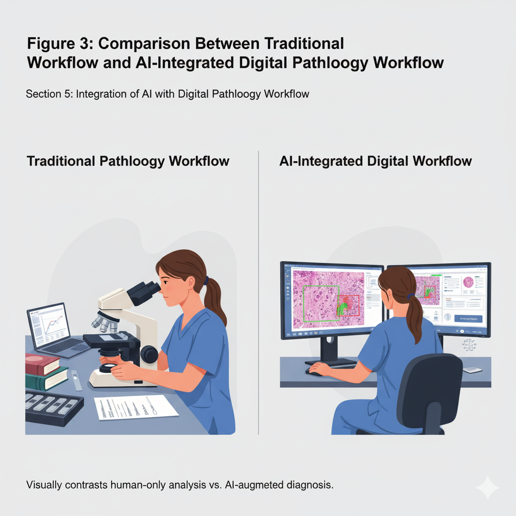
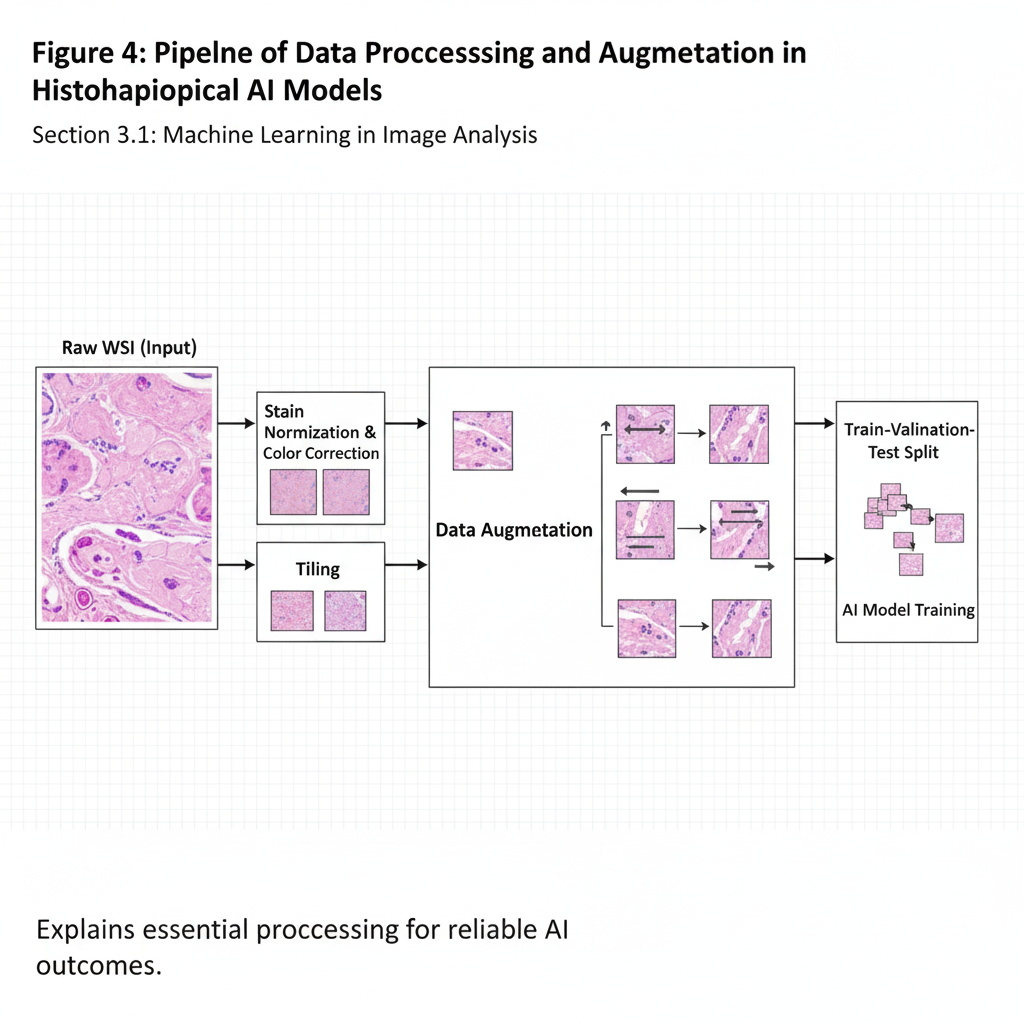
The transfer learning enables the use of models trained on large datasets (such as ImageNet) to fine-tune to perform historical jobs. It saves on training time and enhances accuracy particularly where annotated medical data are not abundant. Even in cases where training is done using small datasets, pretrained CNNs can be useful in identifying morphological patterns (Spasic & Nenadic, 2020).
APPLICATIONS OF AI IN HISTOPATHOLOGICAL IMAGE ANALYSIS
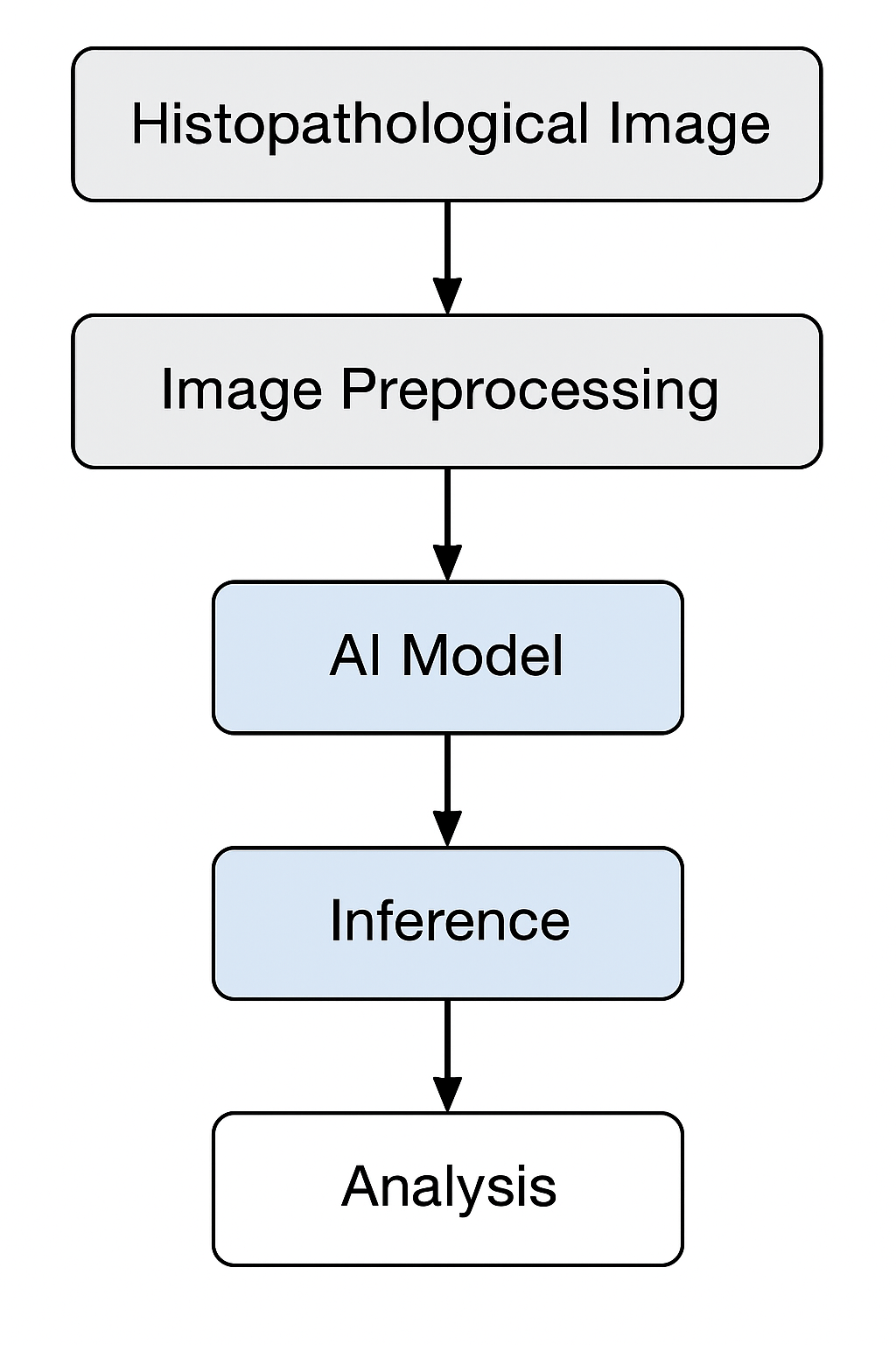
The transformative potential of AI in the histopathology field is in the diagnostic and prognostic fields, as well as in research. The workflow of AI-assisted histopathological image analysis (Fig. 4).
Automated tissue classification: The AIs are capable of identifying normal, benign and malignant tissues with accuracy. To illustrate, CNN-based classifiers have been able to distinguish breast carcinoma and normal tissue with more than 95 percent accuracy (Sampath et al., 2024). Automated classification will help to triage the slides faster and help pathologists to focus on the high-risk cases.
Tumor detection and grading: AI has been found to be very useful in identifying tumor areas and attaching a histological grading (Fig. 5; Table 2). CNNs are able to detect malignant glands in prostate and breast cancers and grade tumors based on some known standards like the Gleason or Nottingham grading systems. AI algorithms decrease the intra-and inter-observer variability and give stable results.
Cell Segmentation and Counting: Precise segmentation of nuclei, cytoplasm, and other structures is crucial for quantitative histopathology. Deep learning-based segmentation algorithms, such as U-Net and Mask R-CNN, outperform traditional thresholding techniques by capturing complex cellular boundaries (Xu et al., 2024). Automated cell counting facilitates objective measurement of mitotic index, necrosis, and immune infiltration.
Prediction of genetic and molecular markers: AI may discern genetic mutation and molecular changes directly based on histological morphology, the principle of which is termed as computational histogenomics. As an example, deep models have predicted microsatellite instability (MSI) and KRAS mutations and HER2 status using H&E slides in colorectal and breast cancers, which may lessen the need to use expensive molecular tests (Guitton et al., 2023).
AI in cancer diagnosis and prognosis: AI-based prognostic models are prognostic models that combine both the histopathological features and clinical data to determine the outcome of the patient. As an example, deep learning devices that analyze lung adenocarcinoma slides have been able to forecast the survival time and risk of recurrence. These models facilitate individualized therapy choices and accuracy in oncology (Kumar, 2022).
| Algorithm | Feature Type | Typical Applications | Advantages | Limitations | Reference |
|---|---|---|---|---|---|
| Support Vector Machine (SVM) | Texture, Color, Shape | Tumor classification, cell type identification | Robust for small datasets | Requires feature engineering | Khan et al., 2023 |
| Random Forest | Morphometric features | Tissue segmentation | Handles nonlinear data | Limited interpretability | Vishraj et al., 2023 |
| K-Nearest Neighbors (KNN) | Spatial features | Cell clustering | Simple implementation | Computationally expensive for large data | Halder et al., 2024 |
| Decision Tree | Statistical features | Region classification | Easy interpretation | Prone to overfitting | Halabaku & Bytyci, 2024 |
| Cancer Type | AI models applied to tumor detection and grading | ||
|---|---|---|---|
| AI Architecture | Accuracy / Performance Metric | Reference Study | |
| Breast cancer | ResNet-50 CNN | 96% classification accuracy | Behar & Shrivastava, 2022 |
| Prostate cancer | Inception-v3 | 93% accuracy in Gleason grading | Murtaza et al., 2025 |
| Lung adenocarcinoma | VGG-16 + Transfer Learning | 94% sensitivity | Humayun et al., 2022 |
| Colorectal carcinoma | Custom CNN | 91% F1-score | Sadagopan et al., 2023 |
| Step | Components of a digital pathology & AI workflow | ||
|---|---|---|---|
| Description | Tools / Techniques | References | |
| Image acquisition | WSI scanners capture digital slides | Leica, Hamamatsu, Aperio | Ardon et al., 2025 |
| Pre-processing | Normalize color, remove blur | Reinhard normalization, CLAHE | Ningsih, 2020 |
| Patch extraction | Divide slide into smaller images | Sliding window, ROI selection | Hossain et al., 2023 |
| Model training | Deep learning on labeled data | TensorFlow, PyTorch | Stevens et al., 2020 |
| Post-processing | Aggregate predictions, generate heatmaps | Grad-CAM, probability mapping | Ariyametkul & Paing, 2025 |
AI in non-cancerous histopathology: In addition to oncology, AI is useful in the diagnosis of inflammatory diseases, fibrosis and metabolic disorders. Stages of liver fibrosis have been classified by automated systems, lesions of diabetic nephropathy have been identified, and immune cell infiltration in autoimmune diseases has been quantified, all showing the universality of AI in pathological fields (Decharatanachart et al., 2021).
INTEGRATION OF AI WITH DIGITAL PATHOLOGY WORKFLOW
The adoption of AI demands powerful WSI systems that can handle big data, which can be many gigabytes of data per slide. AI pipelines consist of preprocessing (color normalization, artifact removal), patch extraction, model training and post-processing. This volume of data needs efficient data storage and retrieval systems (Smith et al., 2021). The cloud computing technology provides mass computation and interaction between institutions. Edge AI Edge computing on devices can decrease data transmission time by processing data on the device, and increase privacy, enabling real-time slide processing in the diagnostic processes (Hossain et al., 2023).
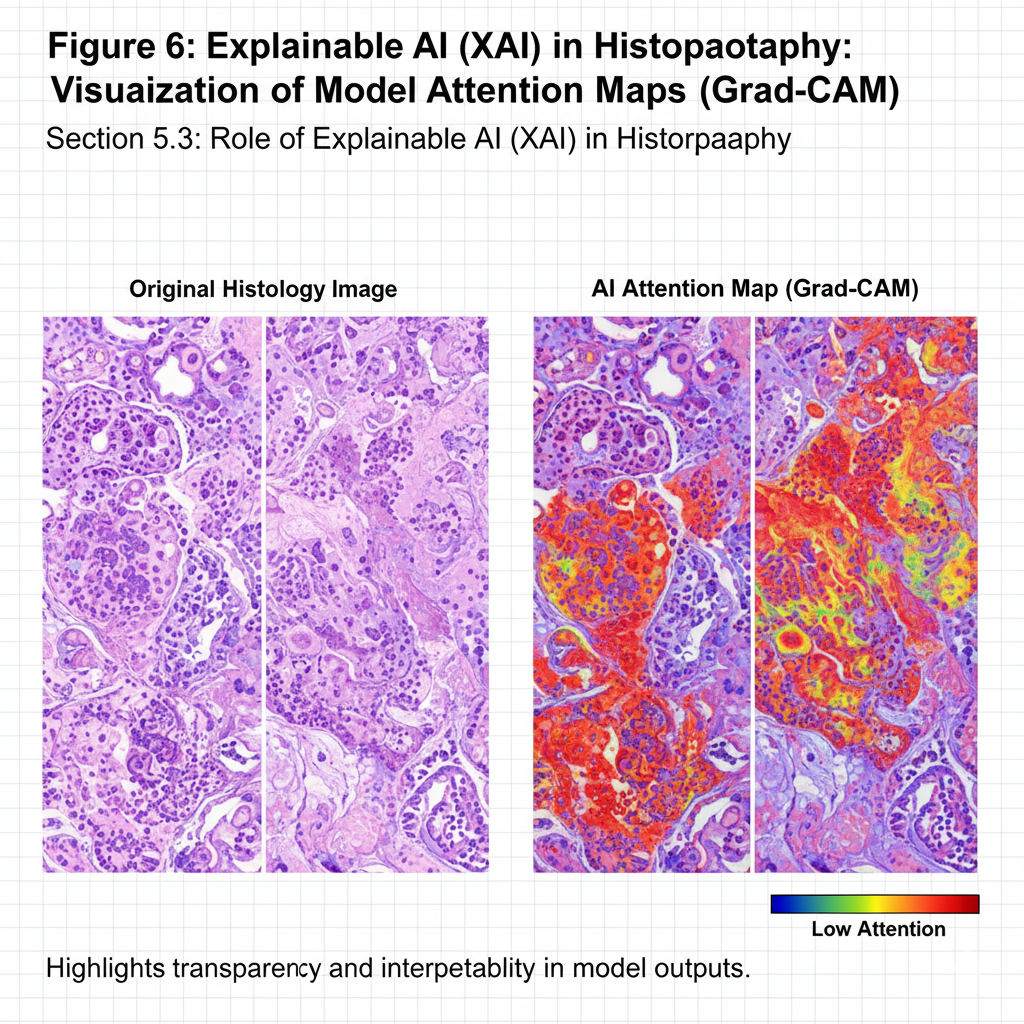
Although AI models can be highly accurate, the lack of transparency reduces their use by clinicians due to their black-box design (Marey et al., 2024). Explainable AI offers visualization tools (e.g., Grad-CAM, heatmaps) to show parts of an image that affect the choice of a model as shown Fig. 6. This openness gives confidence to the pathologists and helps to validate the model. The components of a digital pathology and AI workflow is shown in Table 3.
CHALLENGES AND LIMITATIONS
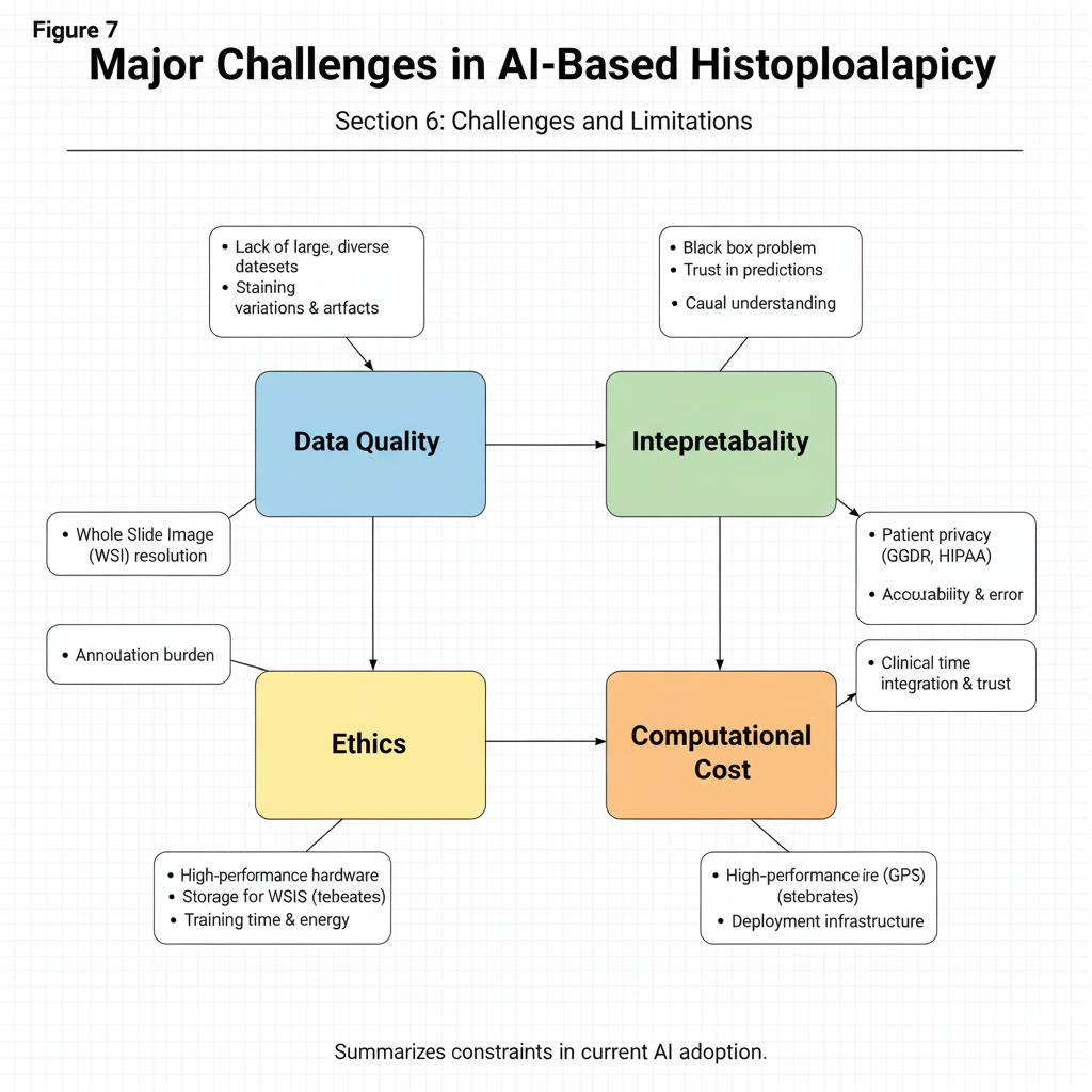
Nevertheless, although there could be impressive advances in AI histopathology, there are a number of technical, computational, ethical, and regulatory challenges that restrict its ubiquitous application in clinical practice (Fig. 7; Table 4). These issues do not only influence the performance and reliability of AI systems but also the acceptance of AI systems by clinicians, regulatory agencies, and patients. To ensure the utilization of AI tools in the pathology practice is both safe, fair, and effective, it is necessary to understand and mitigate these limitations (Ahmad et al., 2021).
Data and Annotation Issues
The quality and availability of annotated data is one of the most basic problems of AI-based histopathology. Training deep learning models that are able to produce accurate and generalizable results requires high-quality and labeled datasets. Nevertheless, data annotation in pathology is a process, which is complex, time-consuming, and subjective. It can be a very tedious task in which thousands of microscopic images have to be manually labeled by expert pathologists, bringing variability in terms of expertise, interpretation and experience (Sultan et al., 2020).
Additionally, histopathological data are usually affected by the lack of consistency due to the variation in staining methods (e.g. hematoxylin and eosin intensities), types of scanners, magnification, and resolutions of images. The result of these inconsistencies is noise and bias, and thus poorer performance of the model in the real-world clinical environments. The other outstanding problem is the non-standardization of different institutions and it is hard to develop strong datasets that reflect different populations and disease conditions. These issues can be overcome by establishing standard data protocols, automated labeling methods and multi-institutional initiatives to generate balanced and representative datasets (Dunn et al., 2025).
Model Interpretability
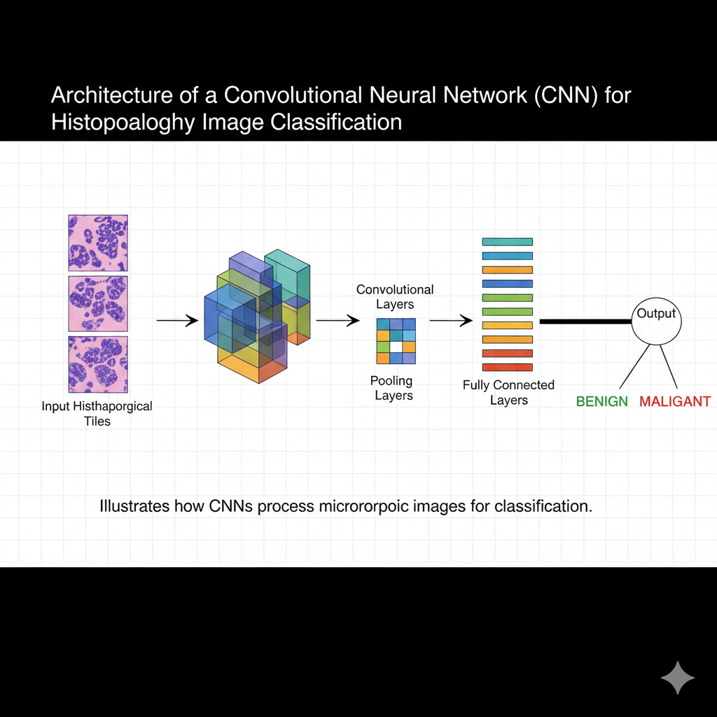
Deep learning models, especially convolutional neural networks (CNNs) get the name black box because they are not very interpretable. Even though such models can be highly accurate in diagnosing activities, they do not often give evidence of the rationale of their forecasts. Pathologists and regulatory authorities in the clinical practice demand clear and understandable outputs as a way of maintaining accountability and trust.
Without interpretability, it is hard to detect possible errors, biases or overfitting in AI predictions. It also prevents clinicians to learn the histological features that have led to a particular diagnosis. To counter this, explainable AI (XAI) methods are under development, including heatmaps, saliency maps, and attention mechanisms that can be used to visualize the decision-making pathway and provide important diagnostic areas in a tissue sample. Nevertheless, striking a balance between the complexity of the model and the interpretability of the result is one of the key research areas in the field of modern computational pathology (Bhati et al., 2024).
Ethical and Regulatory Considerations
The use of AI in medical diagnostics comes with a number of ethical and regulatory issues. The most crucial issue is data privacy and patient confidentiality, histopathological images, and metadata can be used to identify a patient. Furthermore, biases in algorithms due to asymmetric training data result in the inequalities of the performance with various groups of patients that may cause disparities in health (Paulus & Kent, 2020).
The issues of accountability and liability are also complicated: it is not clear who should be held responsible in case of diagnostic errors caused by AI systems. To address these risks, regulatory bodies like the U.S. Food and Drug Administration (FDA) and the European Medicines Agency (EMA) are putting in place systems of the assessment of AI-driven medical equipment. These frameworks are concerned with safety, transparency, reproducibility and post-market surveillance to provide ethical compliance and safety to patients (Al-Dulaimi & Mohammad, 2025).
Computational and Resource Constraints
The second significant obstacle to the implementation of AI in histopathology is that it is computationally expensive to train and deploy deep learning models. Whole-slide images (WSIs) are gigapixel images that demand a lot of memory, storage and high-performance computing infrastructures to process (Ahmed et al., 2022). Not every laboratory, particularly in the low- and middle-income nations, has such sophisticated resources and is thus able to adopt AI systems in practice. Furthermore, the maintenance and update of AI models require constant retraining on new data, which requires constant technical skills and money. The alternative solution is cloud-based AI solutions, which present new issues on data security and local privacy regulations. Therefore, the fair access to the AI-based diagnostic technologies should be provided by working on the creation of computationally efficient algorithms and the resource-sharing models among the research and clinical institutions.
Future Directions
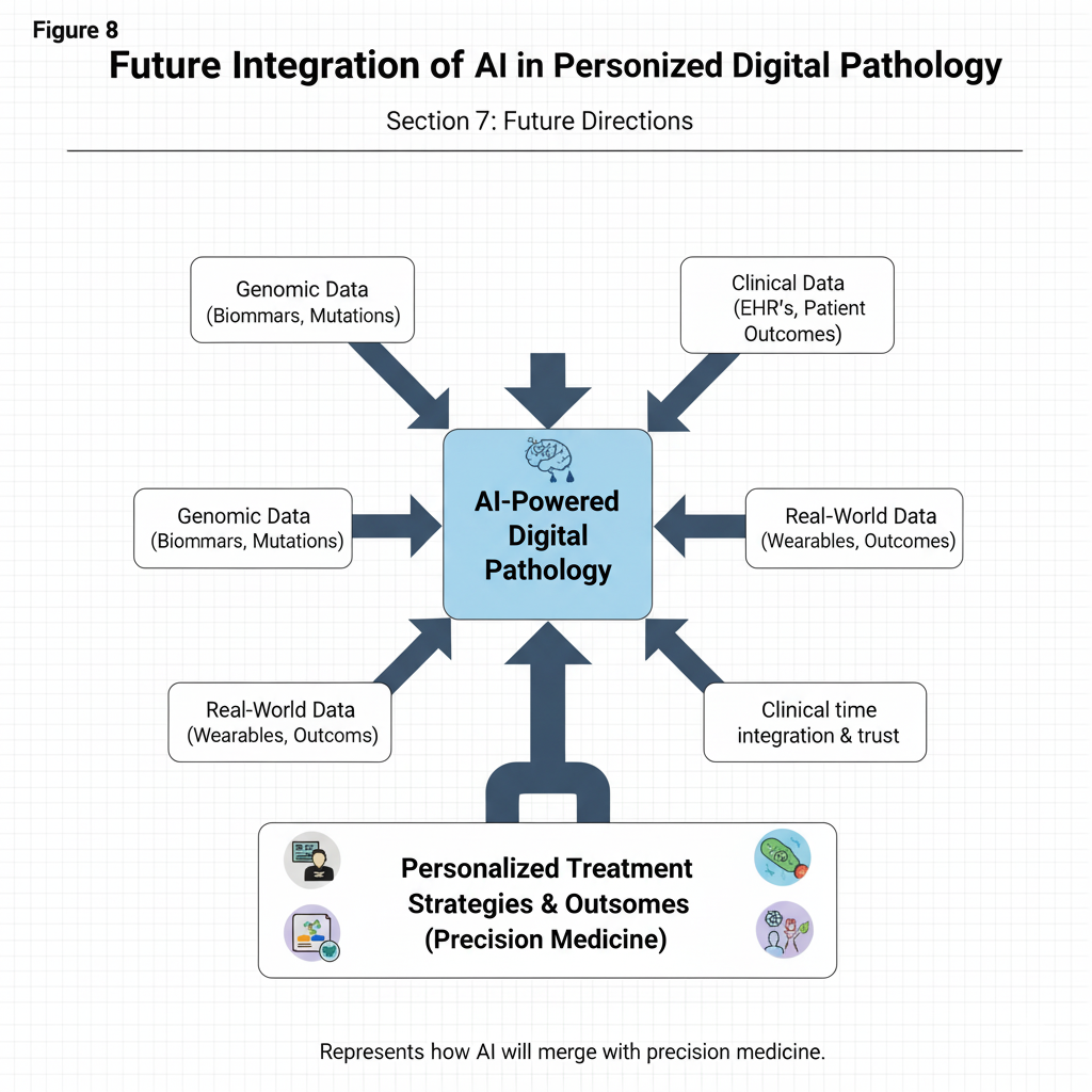
AI is still transforming the face of histopathology and is likely to bring a time when diagnosis, prognosis, and treatment planning is quicker, more accurate, and more personalized as shown in Fig. 8 and Table 5. Although the present AI systems have shown important diagnostic potential, future developments will be aimed at combining other biological information, enhancing human-machines cooperation, providing privacy to data, and giving medicine to individual patients. These guidelines will reinvent the role of histopathology in the field of precision healthcare at large (Lin, H et al., 2024).
Combination with Multi-Omics Data
In the future of AI-based histopathology, the focus will be on combining multi-omics data (genomics, transcriptomics, proteomics, and metabolomics) with digital pathology images. Conventional histopathology is based mostly on morphologic characteristics that can be observed under the microscope, however, the morphologic characteristics may be studied as an indication of underlying molecular changes that are not necessarily visible to the naked eye. AI can detect complicated biological patterns in molecular-level data and predict novel biomarkers of disease progression, therapeutic resistance, or prognosis by integrating molecular level data with tissue morphology (Tang et al., 2022).
As an example, deep learning models are capable of matching gene expression patterns to particular histological buildings, meaning AI can identify molecular subtypes of cancer directly on images. This kind of integration will improve accuracy of diagnosing and clinicians can classify diseases and be more predictive of what to expect of a patient. Also, such a multimodal approach will be able to drive into the identification of new therapeutic targets and provide a gap linking molecular biology and digital pathology, which will result in a more in-depth form of disease mechanisms (Boehm et al., 2022).
AI-Augmented Pathologists
The replacement of human expertise in the field of pathology will not be the future but rather the use of AI-enhanced collaboration. AI will not replace pathologists but will be an aid device, which will complement human decision-making, reduce diagnostic errors, and simplify the efficiency of the working process. The AI systems will automatically identify the abnormal areas, calculate the tissue parameters, and give priority to complex or unclear cases to be examined by experts (Ahmad et al., 2021).
An interdisciplinary strategy will lessen the diagnostic fatigue levels and enable pathologists to concentrate on cases that need high cognitive interpretation. Besides, AI may also serve as a partner of continuous learning through real-time feedback and standardized quality evaluation. Medical education will also use AI-based tools in training settings with virtual slide annotation and automated tests of histopathology trainees. This machine intelligence and human expertise will eventually enhance the accuracy and consistency of diagnosis as well as patient outcomes (Hirosawa & Shimizu, 2025).
Federated Learning in Medical Imaging
| Category | Major challenges in AI-based histopathology | ||
|---|---|---|---|
| Description | Potential Solutions | References | |
| Data variability | Differences in staining, scanners, and slide quality | Color normalization, domain adaptation | Hetz et al., 2024 |
| Limited annotations | Manual labeling is time-consuming | Semi-supervised and weakly supervised learning | Qu et al., 2022 |
| Interpretability | “Black-box” nature of deep models | Explainable AI, saliency mapping | Hassija et al., 2024 |
| Ethical and legal issues | Data privacy, bias, accountability | Secure data sharing, regulatory standards | Renuka et al., 2025 |
| Computational demand | High storage and GPU requirements | Cloud and federated learning frameworks | Albshaier et al., 2025 |
| Area | Emerging research directions in AI-based histopathology | ||
|---|---|---|---|
| Objective | Expected Impact | References | |
| Multi-omics integration | Combine histology with genomic data | Precision diagnosis | Wekesa & Kimwele, 2023 |
| AI-assisted workflows | Support pathologists with automated tools | Reduced workload, improved accuracy | Tariq, 2024 |
| Federated learning | Enable decentralized model training | Data privacy, scalability | Kanji, 2020 |
| Real-time diagnostics | On-scanner inference using Edge AI | Faster turnaround time | Avci et al., 2025 |
| Personalized therapy | Predict treatment response | Tailored patient management | Wei & Chu, 2022 |
Limited access to large, varied, and ethically shareable datasets is one of the greatest constraints to the development of AI. Federated learning is a radical solution as it allows individual hospitals or research centers to exchange AI models without sharing raw patient data. Rather, model parameters are distributed among institutions, thus protecting patient confidentiality and enjoying the benefits of joint learning.
This decentralized model is as much as ensuring that the data privacy rules like GDPR and HIPAA are met but it also enhances the generalization of the model because it will expose the AI to diverse data demographic and technical. With the increased use of federated learning, it will help to develop global AI models in pathology that are strong, objective, and can be used with various populations and imaging systems (Ettaloui et al., 2023).
AI in Personalized Medicine
The incorporation of AI into personalized medicine is one of the most promising areas of digital pathology. Through observing minute morphological changes in tissues and correlating them with clinical results, AI will be able to foresee drug reactions, disease recurrence, and the prognosis. This forecasting ability allows creating the treatment plans that are unique and targeted at addressing the biological characteristics of a particular patient.
As an example, AI models can assist oncologists in determining patients who most likely respond to certain targeted therapy or immunotherapy and reduce needless therapy and side effects. In the long term, AI systems can also facilitate longitudinal monitoring, which evaluates the response of the tissues to the therapy and can change the treatment plans in a dynamic way (Reddy et al., 2021).
CONCLUSION
AI has ushered in a new era in histopathological image analysis, transforming subjective microscopy into an objective, quantifiable science. Through deep learning and advanced computational frameworks, AI can detect tumors, classify tissues, and predict molecular markers with exceptional accuracy. Despite challenges in data quality, interpretability, and ethics, the continuous evolution of AI and digital pathology infrastructure promises a future of more precise, efficient, and accessible diagnostics. The integration of AI into routine pathology practice will not replace human expertise but will empower pathologists to make faster, data-driven, and reproducible decisions, ultimately improving patient care and advancing biomedical research.
References
- Ahmad Z, S Rahim, M Zubair et al., 2021. Artificial intelligence in medicine: applications, future role, obstacles, and philosophical considerations. Diagnostic Pathology 16:24.
- Albshaier L, S Almarri & A Albuali, 2025. Federated learning for cloud and edge security: systematic review. Electronics 14:1019.
- Al-Dulaimi AOM & MAAW Mohammed, 2025. Legal responsibility for AI-caused errors in public sector. International Journal of Law and Management 2025:0295.
- Ardon O, A Manzo, J Spencer et al., 2025. Digital slide scanning at scale: comparison of whole slide imaging devices. Journal of Pathology Informatics 100446.
- Ariyametkul A & MP Paing, 2025. Explainability of YOLO-based breast cancer detection using heatmaps. Quantitative Imaging in Medicine and Surgery 15:6252-71.
- Ataky STM, JD Matos, AS Britto Jr et al., 2021. Machine learning methods for histopathological image analysis: a review. Electronics 10:562.
- Avci MB, F Kurul, SN Topkaya et al., 2025. Smartphone-based biosensing: optical imaging, microfluidics and AI. Microchimica Acta 192:786.
- Bahadir CD, M Omar, J Rosenthal et al., 2024. Artificial intelligence applications in histopathology. Nature Reviews Electrical Engineering 1:93-108.
- Balestrieri A, C Politi, F Coghe et al., 2025. The evolution from qualitative to quantitative analysis in medical sciences: a historical perspective and the impact of AI 11:400-12.
- Behar N & M Shrivastava, 2022. ResNet50-based effective model for breast cancer classification using histopathology images. CMES–Computer Modeling in Engineering and Sciences 130:823-39.
- Bhati D, F Neha & M Amiruzzaman, 2024. Survey on explainable AI techniques for visualizing deep learning models in medical imaging. Journal of Imaging 10:239.
- Biswas B, 2025. Integrative diagnostic pathology: cytomorphology, genomics, and translational perspectives. Deep Science Publishing.
- Boehm KM, P Khosravi, R Vanguri et al., 2022. Multimodal data integration to advance precision oncology. Nature Reviews Cancer 22:114-26.
- Decharatanachart P, R Chaiteerakij, T Tiyarattanachai et al., 2021. Artificial intelligence in non-alcoholic fatty liver disease and fibrosis: systematic review. Therapeutic Advances in Gastroenterology 14:1-17.
- Dika E, N Curti, E Giampieri et al., 2022. Manual and automatic computer-aided vs. traditional histopathological diagnosis of melanoma. Pathology Research and Practice 237:154014.
- Dunn C, D Brettle, C Hodgson et al., 2025. International study of stain variability in histopathology. Journal of Pathology Informatics 17:100423.
- Ettaloui N, S Arezki & T Gadi, 2023. Blockchain-based electronic health records and GDPR/HIPAA compliance. Data and Metadata 2:166.
- Guitton T, P Allaume, N Rabilloud et al., 2023. AI predicting microsatellite instability and KRAS/BRAF mutations. Diagnostics 14:99.
- Gupta E, P Bhalla, N Khurana et al., 2009. Histopathology for diagnosis of infectious diseases. Indian Journal of Medical Microbiology 27:100-6.
- Halabaku E & E Bytyçi, 2024. Overfitting in machine learning: comparative analysis. Intelligent Automation and Soft Computing 39:987-1006.
- Halder RK, MN Uddin, MA Uddin et al., 2024. Enhancing K-nearest neighbor algorithm: review and performance analysis. Journal of Big Data 11:113.
- Hassija V, V Chamola, A Mahapatra et al., 2024. Interpreting black-box models: review on explainable AI. Cognitive Computation 16:45-74.
- Hetz MJ, T-C Bucher & TJ Brinker, 2024. Multi-domain stain normalization in digital pathology. Medical Image Analysis 94:103149.
- Hirosawa T & T Shimizu, 2025. Narrative review of AI in medical diagnostics. Computers, Materials and Continua 83:3919-44.
- Hossain ME, MTR Tarafder, N Ahmed et al., 2023. AI with edge computing for real-time decisions. International Journal of Multidisciplinary Sciences and Arts 2:252-61.
- Hossain MS, GM Shahriar, MMM Syeed et al., 2023. Vision transformer–based ROI selection for WSI. Scientific Reports 13:11314.
- Humayun M, R Sujatha, SN Almuayqil et al., 2022. Transfer learning with CNN for lung carcinoma classification 11:400-12.
- Jain E, A Patel, AV Parwani et al., 2024. Whole slide imaging: current and emerging perspectives. International Journal of Surgical Pathology 32:433-48.
- Kanji RK, 2020. Federated learning in big data analytics. Journal of Scientific and Engineering Research 7:343-52.
- Khan A, A Sohail, U Zahoora et al., 2020. Survey of deep CNN architectures. Artificial Intelligence Review 53:5455-516.
- Khan F, Y Gulzar, S Ayoub et al., 2023. LS-SVM-based brain tumor classification. Frontiers in Applied Mathematics and Statistics 9:1324054.
- Kumar A, 2022. AI in digital pathology for cancer grading. International Journal of Computer Applications Technology Research 11:400-12.
- Lin H, J Han, P Wu, et al., 2024. Machine learning and human-machine trust in healthcare. CAAI Transactions on Intelligence Technology 9:286-302.
- Lowe JS, PG Anderson & SI Anderson, 2023. Stevens & Lowe's Human Histology. Elsevier Health Sciences 09:440-452.
- Marey A, P Arjmand, ADS Alerab et al., 2024. Explainability and black-box challenges in cardiovascular radiology. Egyptian Journal of Radiology and Nuclear Medicine 55:183.
- Murtaza G, R Kausar, S Sial et al., 2025. Targeting diabetic wound healing through nanoparticles in streptozotocin-induced rats. Pakistan Veterinary Journal 45:1244-52.
- Ningsih DR, 2020. Improving retinal image quality using histogram equalization. International Journal of Image, Graphics and Signal Processing 14:30.
- Paulus JK & DM Kent, 2020. Algorithmic prediction and health disparities. NPJ Digital Medicine 3:99.
- Qu L, S Liu, X Liu et al., 2022. Weakly- and self-supervised deep learning for histopathology: survey. Physics in Medicine and Biology 67:20TR01.
- Reddy MS, M Sarisa, S Konkimalla et al., 2021. AI/ML in disease forecasting. ESP Journal of Engineering and Technology Advancements 1:188-200.
- Renuka O, N RadhaKrishnan, BS Priya et al., 2025. Data privacy and protection: legal and ethical challenges. In: Emerging Threats and Countermeasures in Cybersecurity (Shrivastava G, RP Ojha, S Awasthi et al., Eds.): Wiley, Hoboken, USA, pp: 433-65.
- Sadagopan R, S Ravi, SV Adithya et al., 2023. PolyEffNetV1 for colorectal polyp detection. Journal of Engineering in Medicine 237:406-18.
- Sampath K, S Rajagopal & A Chintanpalli, 2024. CNN architectures for early bone cancer diagnosis. Scientific Reports 14:2144.
- Smith B, M Hermsen, E Lesser et al., 2021. Image analysis pipelines for WSIs. Journal of Clinical and Translational Science 5:e38.
- Spasic I & G Nenadic, 2020. Clinical text data in machine learning: systematic review. JMIR Medical Informatics 8:e17984.
- Sultan AS, MA Elgharib, T Tavares et al., 2020. AI, ML and DL in oncologic histopathology. Journal of Oral Pathology and Medicine 49:849-56.
- Tang S, K Yuan & L Chen, 2022. Biomarkers for rare disease diagnosis. Fundamental Research 2:894-902.
- Tariq M, 2024. AI in pathology: diagnostic accuracy and workflow. International Journal of Applied Biology and Forensic 8:141-50.
- Vishraj R, S Gupta & S Singh, 2023. Feature selection for lung tissue categorization using HRCT. Expert Systems 40:e13320.
- Wei M & C-Q Chu, 2022. Predicting treatment response in rheumatoid arthritis. Best Practice and Research Clinical Rheumatology 36:101741.
- Wekesa JS & M Kimwele, 2023. Multi-omics data integration using deep learning. Frontiers in Genetics 14:1199087.
- Xu Y, R Quan, W Xu et al., 2024. Advances in medical image segmentation. Bioengineering 11:1034.